this lecture dealt with some of the higher order processing of the somatosensory cortex; namely, the cortex's ability for localization and discrimination of fine textures and resolutions. localization refers to how accurately the somatosensory cortex can distinguish a stimuli from a certain location on the body. the two point discrimination test measures the smallest distance between two simultaneous stimuli which will indeed produce a sensation of two stimuli.
lateral inhibition is introduced as a means of increasing the resolution and sensitivity to stimuli, a mechanism which is used widely in the cortex, thalamus, and dorsal column nuclei. the neuron at the center of a given receptive field essentially produces the strongest signal because of its inhibition of neighboring nuclei, which are also inhibiting neighboring nuclei. this inhibition occurs via inhibitory interneurons, which are stimulated by recurrent branches of the dorsal column nuclei.
the cortex can also regulate lateral inhibition to enhance the selectivity of the sensory input even further; this is seen especially during movement, where the cortical neurons will directly stimulate or inhibit dorsal column nuclei to select for stimuli that is relevant to the movement, allowing for greater discrimination of complex textures.
the next section looked at the somatosensory association cortex, which lies posterior to the primary somatosensory cortex. it is responsible for deciphering the meaning behind tactile input, formation of an egocentric space via the integration of tactile, proprioceptive, auditory, and visual information, and language comprehension via Wernicke's area. lesions within this area can produce such syndromes as astereognosis, which is the inability to identify an object by touch, or neglect syndrome, which is a loss of cognition in half of the sensory field.
finally, two conditions are discussed related to spinal cord injury: syringomyelia is the formation of cysts in spinal grey matter which blocks decussation of anterolateral second order neurons and thus the bilateral loss of pain and temperature sensation. brown-sequard syndrome results from a hemisection of the spinal cord, an injury that slices through the dorsal and anterolateral columns on one side, that results in loss of ipsilateral motor control and discriminative touch sensation, and loss of contralateral pain and temperature sensation.
questions
localization...
1. cortical localization requires...
2. how are receptive fields differentiated in peripheral structures?
3. what is the relationship between localization and receptive field size?
4. how are receptive fields differentiated in the CNS?
lateral inhibition...
5. what is lateral inhibition?
6. describe what happens with stimulation of the center and peripheral region of a receptive field for a given dorsal column nuclei neuron.
7. what are inhibitory interneurons and what are their function in lateral inhibition?
8. what is the net effect of lateral inhibition?
two point discrimination, cortical regulation...
9. what is meant by "two point discrimination"?
10. describe how two point discrimination is related to localization and lateral inhibition.
11. how does syphilis affect the two point discrimination test?
12. how does the cortex regulate lateral inhibition?
13. what is the purpose of cortical regulation of lateral inhibition?
14. why does somatosensory perception depend on movement?
somatosensory association cortex...
15. somatosensory association cortex lies...
16. what is the somatosensory association cortex involved in?
17. what is the region within the somatosensory cortex involved in language comprehension?
pathologies...
18. what is astereognosis?
19. what is neglect syndrome?
20. most neglect is due to...
21. why do lesions to the left parietal lobe not necessarily lead to neglect syndrome?
22. what is syringomyelia?
23. what is brown-sequard syndrome?
answers
1. differentiation of neural input from receptive fields at many different levels of processing including the dorsal column nuclei, thalamus, and somatosensory cortex.
2. by the specific activity of sensory neurons that innervate each RF.
3. the smaller receptive fields have a greater degree of localization.
4. via neural interactions between pathways between different RF's.
5. a neural mechanism employed in the cortex, thalamus, and dorsal column nuclei that enhances the difference between somatosensory receptive fields.
6. stimulation at the center will produce a stimulatory effect on the dorsal column nuclei neuron and stimulation at the periphery of the receptive field will produce an inhibitory effect.
7. inhibitory interneurons are the neurons that are stimulated by the recurrent branches of dorsal column nuclei which inhibit action of neighboring dorsal column nuclei neurons.
8. lateral inhibition enhances the signal received at the central region of activation and decreases the signal away from the center.
9. the ability to distinguish between separate but simultaneous pinpricks to the skin.
10. lateral inhibition refines the localization of sensory stimulation, which is what is being measured in two point discrimination.
11. the dorsal column degeneration interferes with the reception of discriminative touch and thus reduces one's two point discrimination ability.
12. via descending pathways from the primary somatosensory cortex and motor cortex which modulate afferent inputs.
13. to focus on the afferents that are delivering useful information.
14. during movement, motor cortex sends messages to dorsal column nuclei to facilitate sensory stimuli related to the movement, in a way that increases the discrimination of complex textures.
15. posterior to the primary somatosensory cortex.
16. deciphering meaning of tactile stimuli, integration of touch, proprioception, vision, audition (formation of ego-centric space), language comprehension.
17. Wernicke's area.
18. the inability to identify an object by touch.
19. loss of cognition of half of a sensory field.
20. lesions in the right inferior parietal lobe.
21. the functioning right hemisphere has the capacity to compensate for the left's dysfunction but not vice versa.
22. formation of cysts in the center of the spinal grey area that prevents decussation of second order neurons in the anterolateral system, leading to bilateral loss of pain and temperature sensation.
23. a hemisection of spinal cord which cuts through the dorsal and anterolateral columns on one side, leading to loss of ipsilateral discriminative touch and motor control and loss of contralateral pain and temperature.

Showing posts with label somatosensory. Show all posts
Showing posts with label somatosensory. Show all posts
Monday, April 20, 2009
Saturday, April 18, 2009
organ systems III: somatosensory 2a
this lecture looked at some of the basic ideas involved in the development, structure, and function of the somatosensory cortex. roughly speaking, the somatosensory cortex is a portion of the cerebral cortex that processes sensory information from the body-- touch, pain, temperature, proprioception. the cerebral cortex is the outermost layer of the cerebral hemispheres and is formed from the migration of neural plate cells from the medial and lateral ganglionic eminences to the edges of the cortex via glial cells. the migrating cells self organize into 6 semi-distinct layers which contain each contain different cell types which are specialized for different functions; for example, the stellate cells in layer IV receive information from the thalamus, whereas the pyrimidal cells in layer V project information to the other regions of the CNS. in layer V, there are three types of cells: association cells, which project to the ipsilateral cortex (via the longitudinal, cingulate, uncinate fasciculi) and are involved in integration of information, commisural cells which project to the contralateral cortex, and subcortical cells which project to the basal ganglia, thalamus, brain stem, and spinal cord via the internal capsule.
A few other notes about the structural organization of the cortex: cortical columns are sections of the cortex parallel to the surface which are composed of groupings of neurons of similar modality and receptive field; the phenomenon of somatotopy is the mapping out of the body layout on the surface of the brain. somatotopy is also exhibited in the spinal cord and thalamus, where contiguous portions of these CNS structures represent contiguous portions of the body. however, the mapping of the body on the cortex or thalamus is not necessarily representative of the physical shape of the body; this is demonstrated the unusual proportions of the homunculus, which is the physical representation of the body based on the density of receptor sites in the different areas of the cortex that represent different body parts-- for example, because of the high number of receptor sites in the somatosensory cortex for the lips and hands, these parts are unusually large on the homunculus.
MRI is a tool used to detect brain activity and is useful in pinpointing the brain locations which are specialized to process sensory data from specific body parts. the mechanism works by detecting hydrogen ions: a primary vertical magnetic field aligns the hydrogen ions, and a horizontal pulsed magnetic field causes a vibration in the hydrogen ions, the decay from which can be measured by the fMRI machine. the blood flow to the brain can be measured due to the paramagnetic properties of oxygenated hemoglobin and its effect on the hydrogen ions whose energy is measured.
the primary somatosensory cortex is located in the postcentral gyrus, and primarily receives information from the thalamus, which has nuclei that contain groupings of functionally distinct neurons. specific sensory information is processed in the VPL, VPM, medial and lateral geniculate nuclei, and sent to different areas of the somatosensory cortex based on the particular modality and receptive field of information that they encode. cortical relay nuclei include the VA and VL nuclei, which are involved in the communication between the cerebrum, basal ganglia, and the cerebellum. association nuclei (also called pulvinar nuclei) are involved in integrating information from wide areas of the cortex, and there are also non-specific nuclei which are involved in the reticular system of activation and alertness.
the secondary somatosensory cortex (SII) receives information from the primary and as such is involved in higher somatosensory processing; such as processing of bilateral information or forming 3D representations of single objects from multiple sensory sources. the activity of SII is also interrelated with emotional / motivational state, perhaps because of its relationship with the hippocampus and amygdala.
the last part of the lecture dealt with the dynamic aspects of somatosensory processing. it showed an experiment which mapped out neural activity on the somatosensory cortex of a mouse whisker over time-- showing that stimulation of one whisker caused the receptive field to radiate outward as time passed. another graph showed how the same stimulation can elicit different dynamic responses in different brain areas- some brain areas such as the thalamus and cortex undergoing relatively long periods of oscillation between excitatory and inhibitory states whereas other areas such as the sensory trigeminal nuclei produced a much simpler and shorter response. finally, the concept of neural plasticity was introduced in reference to the ability of the somatotopic map to adapt to experience and learning; areas that correspond to body parts that are more frequently used can expand and take over neighboring areas that are not being used.
questions
development...
1. what part of the neural tube is the cerebral cortex derived from?
2. describe the migration of cells in the formation of the cerebral cortex.
3. describe the formation of distinct layers in the cerebral cortex.
4. which layer are stellate cells in and where do they receive sensory input from?
5. what are the cells in layer V that project information to other CNS regions?
6. describe the role of interneurons in the cerebral cortex.
brodmann's areas...
7. what are brodmann's areas?
8. what are the three cell categories in layer V that project to other CNS regions?
9. describe the association cells in layer V.
10. describe the commisural cells in layer V.
11. describe the subcortical cells in layer V.
neurotransmitters and cortical columns...
12. what are some of the neurotransmitters that cortical cells use?
13. what are the main excitatory and inhibitory neurotransmitters?
14. which neurotransmitters does the reticular formation use?
15. what is a cortical column?
16. multiple columns form...
fMRI...
17. brain imaging is monitored by...
18. MRI is often set to detect...
19. describe the mechanism of detection of an MRI.
20. in an MRI image, the amount of pixels are proportional to...
21. what does fMRI use to measure neurally related blood flow?
22. what is the mechanism for detection of blood oxygen level?
thalamus and primary somatosensory cortex...
23. postcentral gyrus receives...
24. what is the thalamus?
25. what are the different types of nuclei present in the thalamus?
26. what are some examples of sensory nuclei and what do they do?
27. what are some examples of cortical relay nuclei and what do they do?
28. what do association nuclei do? what is another name for them?
29. what do non-specific nuclei of the thalamus do?
30. describe the particular types of somatosensory information encoded in brodmann area 3.
31. sensory input from thalamus to area 3b of SI is processed and...
32. areas 3a and 3b converge onto areas 1 and 2, where neurons...
secondary somatosensory cortex...
33. what type of information does the secondary somatosensory cortex receive?
34. lesion of somatosensory cortex causes...
35. SII response depends on...
36. each level of the somatosensory system generates...
37. describe the movement of the receptive field when the somatosensory cortex of a rodent is stimulated by movement of single whiskers.
integration, dynamic aspects of somatosensory cortex...
38. what is a "homunculus"?
39. what are some areas of high and low densities of sensory receptors?
40. what is "somatotopy"? where else is it exhibited in the body?
41. what does neural plasticity have to do with somatotopy?
answers
1. the rostral part of the neural tube; telencephelon (also derivative of basal ganglia).
2. cells migrate from lateral and medial ganglionic eminences (which becomes the basal ganglia) into the cortical surface, along glial cells.
3. cells organize into 6 relatively distinct layers based upon meaningful spatiotemporal patterns that are projected to other brain structures.
4. layer IV, from the thalamus.
5. pyrimidal cells.
6. interneurons form intricate circuits that generate both excitatory and inhibitory activity.
7. histologically distinct regions of the cerebral cortex.
8. association, commisural, subcortical.
9. axons which project to the ipsilateral cortical regions via the longitudinal, uncinate, cingulate fasciculi. involved in integration of information.
10. axons which project to the contralateral cortical regions.
11. axons which project to the basal ganglia, thalamus, brainstem, and spinal cord via the internal capsule.
12. glutamate, GABA, CCK, VIP, neuropeptide Y, somatostatin, substance P, corticotropin releasing hormone.
13. glutamate is excitatory and GABA is inhibitory.
14. AcH, NE, dopamine, serotonin.
15. a grouping of neurons of the same modality and similar receptive field which form a column on the cerebral cortex perpendicular to the surface of the cortex.
16. an array that will map out different body areas or sensory modalities. (somatotopy)
17. functional magnetic resonance imaging.
18. presence of hydrogen ions.
19. a vertical magnetic field aligns the hydrogen ions, and a briefly applied horizontal magnetic field causes the ions to vibrate. the energy released from the recovery from this vibration is measured in the MRI.
20. the amount of hydrogen ions.
21. BOLD- blood oxygen level detection.
22. oxygenated hemoglobin has more paramagnetic than deoxygenated hemoglobin and thus will affect the hydrogen ions measured by the fMRI differently.
23. touch, proprioceptive, pain, and temperature input via both lemniscal and anterolateral system via the VPL of the thalamus.
24. a pair of oval shaped structures in the diencephelon that contain clusters of nuclei which project axons to the cerebral cortex.
25. specific sensory nuclei, cortical relay nuclei, association nuclei, and non-specific nuclei.
26. VPL, VPM, lateral and medial geniculate nuclei- convey information to different cortical areas based on particular modality of information.
27. VA, VL- they establish the connections between the cortex, basal ganglia, cerebellum.
28. integrate sensory information in wide areas of the cortex. also called pulvinar nuclei.
29. convey activity from reticular formation to wide areas of cortex for attention and wakefulness.
30. brodmann area 3a is proprioception and area 3b is SA and RA cutaneous receptors.
31. projected to adjacent areas of primary somatosensory cortex.
32. respond to more abstract somatosensory aspects such as orientation, motion, spatial arrangement.
33. projections from SI representing multiple sources of contact of a single object, or bilateral input from body.
34. loss of fine localization, texture, pressure.
35. behavioral context, or motivational state, because of their connection with the hippocampus and amygdala.
36. a particular spatiotemporal pattern of neural activity across large populations of neurons.
37. the receptive field starts at a particular point in the somatosensory cortex and radiates outwards over time.
38. the sensory representation of the body based on its representation within the cortex.
39. hands and mouth are high, back is low.
40. the mapping out of body areas on regions of cortical surface. also occurs on the spinal cord and thalamus.
41. neural plasticity is manifested in the dynamic adaptation of somatotopic maps according to different patterns of body movement.
Wednesday, April 15, 2009
organ systems: somatosensory part 1
this lecture was an overview of the different types of sensory receptors and pathways involved in somatosensory reception. the first section introduced the concept of sensory transduction in general: how organisms transduce different forms of energy from the environment such as vibration, heat, light, etc. into neuronal action potentials in order to receive information about the environment which allows them to adapt and survive/reproduce. when a sensory receptor is stimulated, it produces a local graded potential, several of which might add up to elicit an action potential if the threshold is reached.
there are several characteristics that are common to all sensory reception systems; first is that the frequency of the action potentials produced is directly proportional to the intensity of stimulation. the axons that propogate the action potentials have varying degrees of myelination as well, which leads to different conducting velocities for different neurons. measuring the "compound action potential" can differentiate between the different velocities of conduction of different neuron bundles within a nerve- the further away the point of measurement is from the stimulus, the more the different bundles are distinguishable via their velocity.
another way that sensory receptors are distinguished is how they adapt to the stimulus. fast adapting receptors produce action potentials at the beginning and end of the stimulus, and fade quickly in between-- signalling that the event starts and ends. slow adapting receptors produce action potentials all throughout the stimulus, signalling that the event is occuring. the underlying mechanism for this differentiation is through the control of calcium and sodium channel permeability.
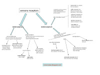 there are a whole variety of particular types of sensory receptors: see chart for more detail. exteroceptors detect outside stimuli and can be divided roughly into two categories: 1) discriminative touch, and 2) non-discriminative touch, pain, and temperature. discriminative touch receptors are covered or modified axonal endings, have faster conducting axons, and project into the CNS via the dorsal lemniscal system (more later). the second category uses free nerve endings as receptors, has slower conducting axons, and project into the CNS using the anterolateral system. interoceptors detect internal stimuli; either from visceral organs or from other internal somatic structures.
there are two distinct pathways by which somatosensory information can travel up to the CNS [see comparison chart]. the dorsal column lemniscal system takes in discriminative touch, proprioception, and some visceral information via first order neurons, which then ascend in the dorsal funiculus, forming the cuneate and gracilis nuclei. in the medulla, they synapse with second order neurons, which decussate (cross over) and form the medial lemniscus, which then travel to the thalamus, where they synapse with the third order neurons, which then project to the somatosensory cortex.
the other pathway for receiving somatosensory input is the anterolateral system, which receives pain, temperature, and non-discriminative touch information. although the distinction between first, second, and third order neurons is not as clear cut as in the lemniscal system, there are still some defining characteristics: first order neurons form vertical tracts of lissaeur, which can span several spinal levels before entering the dorsal horns. first order neurons synapse with second order neurons much lower than in the lemniscal system, which then decussate and project to the anterolateral tract within the lateral funiculi. the second order neurons then project to the thalamus, where they synapse with the lateral thalamic neurons (which project to the primary somatosensory cortex), and the medial thalamic neurons (which project to the cingulate gyrus and insula).
questions
introduction...
1. how is the process of sensation related to survival?
2. describe how our sense organs transduce energy in the environment.
3. sensory receptors are either...
4. what are exteroceptors and interoceptors?
action potentials...
5. describe the production of action potentials by sensory receptors.
6. how does myelination affect the velocity of action potential propagation?
7. what are compound action potentials?
8. how does compound action potential change when the point of measurement is moved further from the point of stimulus?
9. what is the system of nomenclature used to specify motor/sensory nerves within a compound action potential?
10. what are some examples of the assignments of function within the compound action potential naming system?
sensory coding...
11. what are the four attributes of coding that are common to all sensory systems?
12. describe intensity coding of sensory reception.
13. describe the two different mechanisms of "sensory adaption".
14. in general, what is the underlying mechanism for sensory adaptation?
15. what is a cutaneous receptive field?
16. in general, densely innervated RF's are...
17. size and density of RF's provide means for CNS to...
18. what type of proteins are sensory receptor proteins?
exteroception: discriminative touch...
19. describe "discriminative touch".
20. what is the classification (slow adapting or fast adapting) of merkel cells and what are examples of what they are receptive to?
21. ...ruffini corpuscle?
22. ...meissner corpuscle?
23. ...pacinian corpuscle?
24. ...hair follicle?
25. what is a mechanoreceptor receptive field?
26. what is an example of how mechanoreceptor receptive fields aid in forming 3D representations?
exteroception: non-discriminative touch, pain, temperature...
27. what is unique about the sensory reception of non-discriminative touch, pain, and temperature?
28. what is the function and the compound action potential classification of mechanoreceptors for non-discriminative touch?
29. ...thermoreceptors?
30. nociceptors?
31. visceral receptors?
interoception...
32. what are the two types of structures that interoceptors can receive stimulus from?
33. what are visceral afferents?
34. what do visceral nociceptors detect and along which nerves do they travel?
35. describe the reception of pressure, stretch, tension, blood pressure, etc. from visceral organs.
36. what type of sensory information do proprioceptors mediate?
37. what type of receptors are proprioceptors?
38. the muscle receptor proprioceptors detect changes in...
39. the joint receptors detect...
modality...
40. how are the different somatosensory modalities distinguished?
41. describe the characteristics of the discriminative touch, vibration, and proprioception modality.
42. describe the characteristics of the non-discriminative touch, pain, and temperature modality.
43. describe the characteristics of the interoceptive modalities.
different sensory pathways...
44. describe the neuronal pathways that travel through the dorsal lemniscal system and the structure through which the information travels.
45. what are first order neurons?
46. what are second order neurons?
47. what are third order neurons?
48. what modalities travel through the anterolateral system?
49. what is lissauer's tract?
50. what neurotransmitters do first order neurons in the anterolateral system use to synapse with second order neurons?
51. describe the separation of second order neurons by modality within the rexes lamina.
52. where does the neospinothalamic tract project to and what is it involved with?
53. where does the paleospinothalamic tract project to and what is it involved with?
54. where are the third order neurons within the anterolateral pathway and what do they project to?
55. what is the reticular formation?
56. describe the pathway for sensory input and neuronal projection out of the reticular formation.
57. what is a unique anatomical characteristic of reticular formation neurons?
58. what is the physiological effect of the ascending reticular system?
answers
1. the survival of the organism depends on the organism having adequate information about its environment which is gleans via the sensory mechanisms; which provide information that allows the organism to adapt its behavior so as to facilitate survival.
2. our sense organs transduce energy in the form of light, pressure, vibration, sound, etc from the environment into neuronal signals.
3. modified non-neural tissue cells or axons themselves.
4. exteroceptors are sensory receptors that respond to external stimuli such as touch, pain, temperature. interoceptors are sensory receptors that respond to internal stimuli such as chemical change or tissue stretch.
5. stimulation of sensory receptors creates local graded potentials which translate into an action potential which propagates down the axon if the threshold is reached.
6. myelination increases the velocity of action potential propagation.
7. compound action potentials are the sum total of all neuronal activity at a given point due to one stimulation.
8. the further away from the site of stimulation that the compound action potential is measured, the more distinct peaks will be measured based upon velocity of conduction: because of the discrepancy of different axonal clusters based on myelination level and therefore velocity.
9. roman numerals for motor nerves and letters for sensory nerves. lower alphabet letters or lower numbers designate a faster conduction rate.
10. prioprioception and motor neurons are A-alpha. light touch are A-beta. fast pain is A-gamma. slow pain is C.
11. intensity, sensory adaptation, localization, modality.
12. increased intensity of sensory reception increases the frequency of action potential production.
13. rapid adapting receptors respond to stimuli with action potentials that rapidly fade in frequency after the onset or offset of stimuli, indicating that an event occured. slow adapting receptors respond with action potentials that are sustained through the stimuli; indicating that the event is still occuring.
14. calcium or sodium channel activation/inactivation.
15. an area of skin that is innervated by axonal branches off a single neuron.
16. smaller in area.
17. determine location of stimulus on body, distinguish size and shape of stimulus, and resolve spatial resolution.
18. they are transient receptor proteins: each protein responds maximally to a different type of stimulus.
19. a venue of exteroception which is mediated by fast or slow adaptive mechanoreceptors in the CT or around hairs in the epidermal layer.
20. SA1- form and texture, such as fingers scanning a surface. (a murky slow scanner)
21. SA2- skin stretch- perception of hand shape and position. (a dog doing slow taichi)
22. FA- skin movement and slip- for grip control. (mice quickly grip)
23. FA- vibration. (pacman vibrating fast)
24. motion/direction of tactile stimuli. (hair directing a fast picture)
25. a sensory receptive field which contains mechanoreceptors that branch off of a single neuron.
26. the CNS can form a 3D representation of an object held in the hands through the combined contributions of the distinct mechanoreceptive fields from separate digits.
27. they are all mediated by free nerve endings, have slower conducting, A-gamma or C type axons, and are slow adapting.
28. tap, squeeze, rub, skin stretch function, with A-gamma and C axons.
29. hot or cold sensation, A-gamma and C axons.
30. mechano-thermal: mechanical or thermal tissue damage sensation, A-gamma axon. polymodal nociceptor: heat, chemical, tissue damage, C axon.
31. nociception (sympathetic), physiological (parasympathetic), C axon.
32. visceral structures or somatic structures such as the muscles and CT.
33. the afferent nerve fibers that have free nerve ending receptors that mediate pain, pressure, temperature, chemical, and stretch reception in organs and blood vessels.
34. tissue damage or irritation; sympathetic nerves.
35. takes place via physiologic or specialized receptors in smooth muscle, mucosae, hypothalamus- axons travel with parasympathetics.
36. muscle and joint position / movement.
37. fast conducting/large diameter axons, A-alpha,beta
38. length with muscle spindle receptors, and tension with golgi tendon organs.
39. stretch of CT with pacinian and ruffini receptors.
40. through different receptors, conduction velocity / axonal thickness, and the location of their ascending pathway.
41. a more quantitative, localized modality, with sensitive mechanoreceptors, rapid AP conduction, and axons that travel through the lemniscal system within the dorsal column.
42. a more qualitative modality that involves free nerve endings, and axons with slower AP conduction that travel through the anterolateral system.
43. proprioception ascends in the dorsal columns, while visceral sensory information ascends in both dorsal column and anterolateral systems.
44. the dorsal lemniscal system conveys touch, vibration, proprioception, and some visceral information to the cortex by means of first, second, and third order neurons which synapse in the spinal cord, thalamus, and medulla.
45. sensory neurons that form dorsal columns, then synapse onto the gracilis and cuneate nuclei in the medulla.
46. neurons that synapse with first order neurons in the gracilis and cuneate nuclei in the medulla, which then decussate, and ascend to the thalamus, where they synapse with third order neurons.
47. the neurons that synapse with second order neurons in the thalamus and project to the primary somatosensory cortex into the post central gyrus.
48. non-discriminative touch, pain, temperature
49. the ascending and descending of first order neurons within the dorsolateral fasciculus before entering several spinal levels of the gray matter.
50. substance P, glutamate, NO.
51. lamina I and II are associated with pain signals and lamina IV is associated with touch sensation.
52. lateral thalamus and somatosensory cortex, involved in localization of sensation.
53. reticular formation, medial thalamus and cortex- involved in qualitative aspects of pain, temperature, non-discriminative touch.
54. the third order neurons in the lateral thalamus project to the primary somatosensory cortex and the neurons in the medial thalamus project to the cingulate gyrus and insula.
55. a collection of nuclei within the medulla, pons, and midbrain that projects to the cortex and thalamus and is involved in a variety of physiological functions such as alertness, wakefulness, attention.
56. sensory input is received through the raphe and lateral nuclei and is projected upwards through the medial nuclei into the cortex, as well as down into the spinal cord.
57. reticular formation neurons have particularly long axons which allow them to exert an influence over a large area of brain structures.
58. the ascending reticular formation generates alertness and wakefulness.
there are several characteristics that are common to all sensory reception systems; first is that the frequency of the action potentials produced is directly proportional to the intensity of stimulation. the axons that propogate the action potentials have varying degrees of myelination as well, which leads to different conducting velocities for different neurons. measuring the "compound action potential" can differentiate between the different velocities of conduction of different neuron bundles within a nerve- the further away the point of measurement is from the stimulus, the more the different bundles are distinguishable via their velocity.
another way that sensory receptors are distinguished is how they adapt to the stimulus. fast adapting receptors produce action potentials at the beginning and end of the stimulus, and fade quickly in between-- signalling that the event starts and ends. slow adapting receptors produce action potentials all throughout the stimulus, signalling that the event is occuring. the underlying mechanism for this differentiation is through the control of calcium and sodium channel permeability.
 there are a whole variety of particular types of sensory receptors: see chart for more detail. exteroceptors detect outside stimuli and can be divided roughly into two categories: 1) discriminative touch, and 2) non-discriminative touch, pain, and temperature. discriminative touch receptors are covered or modified axonal endings, have faster conducting axons, and project into the CNS via the dorsal lemniscal system (more later). the second category uses free nerve endings as receptors, has slower conducting axons, and project into the CNS using the anterolateral system. interoceptors detect internal stimuli; either from visceral organs or from other internal somatic structures.
there are two distinct pathways by which somatosensory information can travel up to the CNS [see comparison chart].
 the dorsal column lemniscal system takes in discriminative touch, proprioception, and some visceral information via first order neurons, which then ascend in the dorsal funiculus, forming the cuneate and gracilis nuclei. in the medulla, they synapse with second order neurons, which decussate (cross over) and form the medial lemniscus, which then travel to the thalamus, where they synapse with the third order neurons, which then project to the somatosensory cortex.
the other pathway for receiving somatosensory input is the anterolateral system, which receives pain, temperature, and non-discriminative touch information. although the distinction between first, second, and third order neurons is not as clear cut as in the lemniscal system, there are still some defining characteristics: first order neurons form vertical tracts of lissaeur, which can span several spinal levels before entering the dorsal horns. first order neurons synapse with second order neurons much lower than in the lemniscal system, which then decussate and project to the anterolateral tract within the lateral funiculi. the second order neurons then project to the thalamus, where they synapse with the lateral thalamic neurons (which project to the primary somatosensory cortex), and the medial thalamic neurons (which project to the cingulate gyrus and insula).
questions
introduction...
1. how is the process of sensation related to survival?
2. describe how our sense organs transduce energy in the environment.
3. sensory receptors are either...
4. what are exteroceptors and interoceptors?
action potentials...
5. describe the production of action potentials by sensory receptors.
6. how does myelination affect the velocity of action potential propagation?
7. what are compound action potentials?
8. how does compound action potential change when the point of measurement is moved further from the point of stimulus?
9. what is the system of nomenclature used to specify motor/sensory nerves within a compound action potential?
10. what are some examples of the assignments of function within the compound action potential naming system?
sensory coding...
11. what are the four attributes of coding that are common to all sensory systems?
12. describe intensity coding of sensory reception.
13. describe the two different mechanisms of "sensory adaption".
14. in general, what is the underlying mechanism for sensory adaptation?
15. what is a cutaneous receptive field?
16. in general, densely innervated RF's are...
17. size and density of RF's provide means for CNS to...
18. what type of proteins are sensory receptor proteins?
exteroception: discriminative touch...
19. describe "discriminative touch".
20. what is the classification (slow adapting or fast adapting) of merkel cells and what are examples of what they are receptive to?
21. ...ruffini corpuscle?
22. ...meissner corpuscle?
23. ...pacinian corpuscle?
24. ...hair follicle?
25. what is a mechanoreceptor receptive field?
26. what is an example of how mechanoreceptor receptive fields aid in forming 3D representations?
exteroception: non-discriminative touch, pain, temperature...
27. what is unique about the sensory reception of non-discriminative touch, pain, and temperature?
28. what is the function and the compound action potential classification of mechanoreceptors for non-discriminative touch?
29. ...thermoreceptors?
30. nociceptors?
31. visceral receptors?
interoception...
32. what are the two types of structures that interoceptors can receive stimulus from?
33. what are visceral afferents?
34. what do visceral nociceptors detect and along which nerves do they travel?
35. describe the reception of pressure, stretch, tension, blood pressure, etc. from visceral organs.
36. what type of sensory information do proprioceptors mediate?
37. what type of receptors are proprioceptors?
38. the muscle receptor proprioceptors detect changes in...
39. the joint receptors detect...
modality...
40. how are the different somatosensory modalities distinguished?
41. describe the characteristics of the discriminative touch, vibration, and proprioception modality.
42. describe the characteristics of the non-discriminative touch, pain, and temperature modality.
43. describe the characteristics of the interoceptive modalities.
different sensory pathways...
44. describe the neuronal pathways that travel through the dorsal lemniscal system and the structure through which the information travels.
45. what are first order neurons?
46. what are second order neurons?
47. what are third order neurons?
48. what modalities travel through the anterolateral system?
49. what is lissauer's tract?
50. what neurotransmitters do first order neurons in the anterolateral system use to synapse with second order neurons?
51. describe the separation of second order neurons by modality within the rexes lamina.
52. where does the neospinothalamic tract project to and what is it involved with?
53. where does the paleospinothalamic tract project to and what is it involved with?
54. where are the third order neurons within the anterolateral pathway and what do they project to?
55. what is the reticular formation?
56. describe the pathway for sensory input and neuronal projection out of the reticular formation.
57. what is a unique anatomical characteristic of reticular formation neurons?
58. what is the physiological effect of the ascending reticular system?
answers
1. the survival of the organism depends on the organism having adequate information about its environment which is gleans via the sensory mechanisms; which provide information that allows the organism to adapt its behavior so as to facilitate survival.
2. our sense organs transduce energy in the form of light, pressure, vibration, sound, etc from the environment into neuronal signals.
3. modified non-neural tissue cells or axons themselves.
4. exteroceptors are sensory receptors that respond to external stimuli such as touch, pain, temperature. interoceptors are sensory receptors that respond to internal stimuli such as chemical change or tissue stretch.
5. stimulation of sensory receptors creates local graded potentials which translate into an action potential which propagates down the axon if the threshold is reached.
6. myelination increases the velocity of action potential propagation.
7. compound action potentials are the sum total of all neuronal activity at a given point due to one stimulation.
8. the further away from the site of stimulation that the compound action potential is measured, the more distinct peaks will be measured based upon velocity of conduction: because of the discrepancy of different axonal clusters based on myelination level and therefore velocity.
9. roman numerals for motor nerves and letters for sensory nerves. lower alphabet letters or lower numbers designate a faster conduction rate.
10. prioprioception and motor neurons are A-alpha. light touch are A-beta. fast pain is A-gamma. slow pain is C.
11. intensity, sensory adaptation, localization, modality.
12. increased intensity of sensory reception increases the frequency of action potential production.
13. rapid adapting receptors respond to stimuli with action potentials that rapidly fade in frequency after the onset or offset of stimuli, indicating that an event occured. slow adapting receptors respond with action potentials that are sustained through the stimuli; indicating that the event is still occuring.
14. calcium or sodium channel activation/inactivation.
15. an area of skin that is innervated by axonal branches off a single neuron.
16. smaller in area.
17. determine location of stimulus on body, distinguish size and shape of stimulus, and resolve spatial resolution.
18. they are transient receptor proteins: each protein responds maximally to a different type of stimulus.
19. a venue of exteroception which is mediated by fast or slow adaptive mechanoreceptors in the CT or around hairs in the epidermal layer.
20. SA1- form and texture, such as fingers scanning a surface. (a murky slow scanner)
21. SA2- skin stretch- perception of hand shape and position. (a dog doing slow taichi)
22. FA- skin movement and slip- for grip control. (mice quickly grip)
23. FA- vibration. (pacman vibrating fast)
24. motion/direction of tactile stimuli. (hair directing a fast picture)
25. a sensory receptive field which contains mechanoreceptors that branch off of a single neuron.
26. the CNS can form a 3D representation of an object held in the hands through the combined contributions of the distinct mechanoreceptive fields from separate digits.
27. they are all mediated by free nerve endings, have slower conducting, A-gamma or C type axons, and are slow adapting.
28. tap, squeeze, rub, skin stretch function, with A-gamma and C axons.
29. hot or cold sensation, A-gamma and C axons.
30. mechano-thermal: mechanical or thermal tissue damage sensation, A-gamma axon. polymodal nociceptor: heat, chemical, tissue damage, C axon.
31. nociception (sympathetic), physiological (parasympathetic), C axon.
32. visceral structures or somatic structures such as the muscles and CT.
33. the afferent nerve fibers that have free nerve ending receptors that mediate pain, pressure, temperature, chemical, and stretch reception in organs and blood vessels.
34. tissue damage or irritation; sympathetic nerves.
35. takes place via physiologic or specialized receptors in smooth muscle, mucosae, hypothalamus- axons travel with parasympathetics.
36. muscle and joint position / movement.
37. fast conducting/large diameter axons, A-alpha,beta
38. length with muscle spindle receptors, and tension with golgi tendon organs.
39. stretch of CT with pacinian and ruffini receptors.
40. through different receptors, conduction velocity / axonal thickness, and the location of their ascending pathway.
41. a more quantitative, localized modality, with sensitive mechanoreceptors, rapid AP conduction, and axons that travel through the lemniscal system within the dorsal column.
42. a more qualitative modality that involves free nerve endings, and axons with slower AP conduction that travel through the anterolateral system.
43. proprioception ascends in the dorsal columns, while visceral sensory information ascends in both dorsal column and anterolateral systems.
44. the dorsal lemniscal system conveys touch, vibration, proprioception, and some visceral information to the cortex by means of first, second, and third order neurons which synapse in the spinal cord, thalamus, and medulla.
45. sensory neurons that form dorsal columns, then synapse onto the gracilis and cuneate nuclei in the medulla.
46. neurons that synapse with first order neurons in the gracilis and cuneate nuclei in the medulla, which then decussate, and ascend to the thalamus, where they synapse with third order neurons.
47. the neurons that synapse with second order neurons in the thalamus and project to the primary somatosensory cortex into the post central gyrus.
48. non-discriminative touch, pain, temperature
49. the ascending and descending of first order neurons within the dorsolateral fasciculus before entering several spinal levels of the gray matter.
50. substance P, glutamate, NO.
51. lamina I and II are associated with pain signals and lamina IV is associated with touch sensation.
52. lateral thalamus and somatosensory cortex, involved in localization of sensation.
53. reticular formation, medial thalamus and cortex- involved in qualitative aspects of pain, temperature, non-discriminative touch.
54. the third order neurons in the lateral thalamus project to the primary somatosensory cortex and the neurons in the medial thalamus project to the cingulate gyrus and insula.
55. a collection of nuclei within the medulla, pons, and midbrain that projects to the cortex and thalamus and is involved in a variety of physiological functions such as alertness, wakefulness, attention.
56. sensory input is received through the raphe and lateral nuclei and is projected upwards through the medial nuclei into the cortex, as well as down into the spinal cord.
57. reticular formation neurons have particularly long axons which allow them to exert an influence over a large area of brain structures.
58. the ascending reticular formation generates alertness and wakefulness.
Subscribe to:
Posts (Atom)